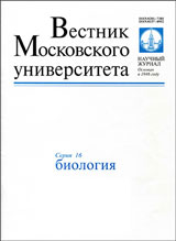
Scientific peer-reviewed journal
"Vestnik Moskovskogo universiteta. Seriya 16. Biologiya" ("Herald of Moscow University. Series 16. Biology") is a peer-reviewed scientific journal of the M.V. Lomonosov Moscow University (School of Biology). Its range includes information on all aspects of modern biology as well as current research on biochemistry, physiology, botany, zoology, anthropology, evolutionary biology, genetics, biophysics, microbiology, developmental biology, gerontology, ecology, etc. The journal is included in the list of the leading periodicals recommended for publishing doctoral research results by the Higher Attestation Commission of the Ministry of Science and Education of the Russian Federation. Since 2015 "Vestnik Moskovskogo universiteta. Seriya 16. Biologiya" is present in Russian Science Citation Index base on the Web of Science platform.
For many years, Moscow University Biological Sciences Bulletin – MUBSB (English edition of "Vestnik Moskovskogo universiteta. Seriya 16. Biologiya" has been published by the Allerton Press (American publishing house), a member since 2007 of the Nauka/Interperiodica International Academic Publishing Company formed in 1992. Since 2007, MUBSB has been distributed by the internationally renowned company Springer Science + Business Media (which is also known as Springer Verlag or simply Springer) according to the contract between this consortium and Pleiades Publishing, Inc. Since 2011, MUBSB is also present in one of the most well-known databases of this type—Scopus. This electronic resource contains both separate papers from the journal and its own page with all scientometric indices. MUBSB also has its own page on the widely renowned SCImago Journal & Country Rank scientific portal. Also MUBSB is indexed in many international databases: EBSCO Discovery Service, OCLC WorldCat Discovery Service, ProQuest Central, AGRICOLA, EMBiology, Institute of Scientific and Technical Information of China, Japanese Science and Technology Agency (JST), Dimensions, Google Scholar etc.
Both journals ' volumes consist of 4 annual issues.
Current issue
Обзоры
This review systematizes contemporary data on the signaling role of mitochondrial metabolites in regulating mitochondrial homeostasis, with an emphasis on their influence on cellular adaptation to stress factors, including age-related changes. Mitochondria function not only as energy sources but also as key sensors and regulators, mediating anterograde and retrograde signaling through metabolites such as citrate, succinate, lactate, and L-carnitine. Citrate and succinate participate in epigenetic modifications, including protein acetylation and succinylation, thereby influencing gene expression and metabolic adaptation, with potential applications in the therapy of oncological and age-associated diseases. Lactate, traditionally regarded as a product of anaerobic metabolism, acts as a signaling molecule that modulates receptor cascades (GPR81), histone lactylation, and oxidative phosphorylation in mitochondria. L-Carnitine ensures metabolic flexibility by maintaining the acyl-CoA/CoA balance, removing toxic metabolites, and enhancing nitric oxide bioavailability, demonstrating its protective effects in models of hyperhomocysteinemia and NO deficiency. Understanding these mechanisms opens up prospects for the identification of biomarkers of mitochondrial dysfunction and the development of therapeutic strategies aimed at restoring homeostasis in the context of aging, metabolic disorders, and gerontological syndromes.
RESEARCH ARTICLES
This article presents the results of a study of the species diversity of representatives of the clade Chlorella in the lake Otstoynik (Tolyatti, Samara region). This reservoir was used until 1996 for the disposal of nitrogen-tuck production waste but is currently at the stage of self-healing. In the course of the work, 15 strains of microalgae with Chlorella-like morphology were studied. Based on the results of molecular genetic analysis using the internal transcribed spacers ITS1 and ITS2, it was found that only 3 strains were true representatives of the genus Chlorella. Microalgae from the genera Brachionococcus, Lobosphaeropsis, Micractinium, and Meyerella were also found. In addition, 2 strains belonged to species that are still formally classified as Chlorella, but their actual taxonomic status needs to be clarified. This study has once again clearly shown that valid identification of Chlorella-like microalgae is not possible using only light microscopy methods. Methods of molecular genetic analysis must be used to study the true species’ richness.
Ulcerative colitis is a socially significant disease, but its etiology is unclear. The most widely used experimental model is dextran sodium sulfate (DSS)-induced colitis. The aim of the work was to characterize morphofunctional and molecular biological changes in the colon and mesenteric lymph nodes in acute colitis induced by 1% DSS solution in male C57BL/6 mice. Upon induction of colitis, moderate ulcerative inflammatory process developed in the colon, hyperplasia of the cortex, plasmatization of the medullary cords and macrophage reaction in the sinuses were observed in the mesenteric lymph nodes. Inflammatory infiltration, increased macrophage content, decreased volume fraction of goblet cells and neutral mucin content in them, increased endocrine cell content, increased expression of Cldn4, Cldn7, Bax and Bcl2 were detected in the colon. Pronounced changes in the composition of intestinal microflora were observed.
The comparative electrophysiological study of auditory sensitivity of intact Wistar rats and heterozygous rats of the transgenic DAT-het line with a reduced expression level of the Slc6a3 gene encoding the dopamine reuptake transporter (DAT-1), providing an experimental model of dopaminergic neurons pathology, was performed. The amplitude and time parameters of auditory brainstem responses recorded in rats of both lines under presentation of paired clicks and single tones were analyzed. Generally, the auditory sensitivities of Wistar and DAT-het rats were similar, but a comparative analysis of the amplitudes of auditory brainstem response peaks, obtained from experimental animals, revealed a significantly greater amplitude of peak 1 in DAT-het rats. Thus, the data obtained suggest that DAT-het heterozygotes differ from rats with a normally functioning dopamine reuptake DAT-1 transporter by an increased level of dopaminergic signaling via activation of D1 receptors localized in the membranes of neurons of the cochlea spiral ganglion.
This article provides a comprehensive study conducted within the Saltykovsky Forest Park, located in a densely urbanised zone east of Moscow. We applied multidisciplinary approach that combines methods of vegetation science, palaeoecology, and historical ecology. Our results indicate that the current flora within the protected natural area exhibits high species richness and contains a relatively low proportion of non-native (adventive) species compared to urban forests in Moscow. Pollen analysis of peat cores extracted from a forest mire enabled the reconstruction of vegetation dynamics spanning the last 4,200 years. The study identified the composition and structure of primary broadleaf forests, phases of agricultural land use, and the period of presentday forests formation. Evidence suggests that initial human activity in the watershed dates back to the Bronze Age, with intensive land use peaking between the 16th and 18th centuries. The oldest part of the park (sectors 1, 3, and 4) encompass ecosystems that have developed over the past 200–300 years on former agricultural land and have undergone multiple stages of ecological succession. Currently, their floristic characteristics closely resemble those of undisturbed zonal forest communities typical of the region. This research underscores the ecological and cultural value of the Saltykovsky Forest Park, recognising it not only as a historic landscape feature but also as a biodiversity refuge that conserves remnants of forest communities typical of the southern taiga zone, despite being embedded in an urban area.
Pulmonary surfactant is an essential component of the respiratory system for the implementation of phagocytosis by alveolar macrophages. Pulmonary tuberculosis is associated with the reduced pulmonary surfactant production and the phagocytic function of macrophages. The transporter protein P-gp (ABCB1 gene) is overexpressed in lung cells and exports numerous substrates. The incorporation of P-gp into the plasma membrane alters its characteristics. The aim of this study was to analyze the relationship between P-gp and the phagocytic activity of macrophages under the influence of exogenous pulmonary surfactant. The study employed scanning electron microscopy and confocal laser scanning microscopy methods, as well as two models of cultured human cells: (1) pro-inflammatory THP-1 macrophages (P-gp+), infected with M. bovis BCG (this model, when exposed to surfactant, is considered a model of alveolarlike macrophages); and (2) parental myeloblastic leukemia K562 cells (P-gp-) and K562/i-S9 cells (P-gp+) transfected with the ABCB1 gene and induced to adhere. In model 1, it was found that the addition of 1 mg/ml of exogenous pulmonary surfactant for 1 h led to the formation of numerous long filopodia, ruffles, and phagocytic cups, as well as a 1.7-fold increase in the phagocytic index. This demonstrates that the surfactant is an effective activator of phagocytosis in infected macrophages. In model 2, it was shown that in the presence of P-gp, the surface activity of cells significantly increased under the influence of exogenous pulmonary surfactant compared to cells without P-gp. It is hypothesized that due to the interaction between P-gp, ERM complex proteins (ezrin, radixin, moesin) and actin filaments, P-gp+ cells are more potentiated for cell surface activation and phagocytosis than P-gpcells. Further analysis of the features of infected macrophages’ phagocytosis depending on P-gp activity may contribute to the development of new drugs aimed at regulating the phagocytic activity of macrophages.
The study of microalgae cell responses to stress factors such as mineral nutrient deficiency is an important ecological task. Changes in the photosynthetic apparatus are reflected in the kinetics of experimentally measured chlorophyll fluorescence induction curves. Various mathematical methods are developed to analyze changes in the shape of curves, allowing rapid analysis of a large number of curves. The paper demonstrates the use of a simple mathematical model of photosystem II (PSII) to assess changes in the PSII parameters of a Chlorella vulgaris microalgae cell culture growing under nitrogen deficiency in the medium. The model describes transitions between three key PSII states that differ in the oxidation state of its components. The mathematical model revealed an increase in the proportion of reaction centers containing smaller antennae, as well as an increase in the proportion of inactive oxygen-releasing complexes.
The data of modern neurobiology indicates a critical dependence of the nervous system formation upon the conditions of intrauterine development. Pregnancy, childbirth and the early postnatal period are of key importance for normal maturation of the nervous system. The developing fetus is especially vulnerable to the effects of adverse external and internal factors in periods of brain and neuronal structures morphological differentiation, during childbirth and the transition to independent breathing. Fetal and/or newborn hypoxia is considered one of the main causes of disorders in brain development, manifested later in form of cognitive impairments, problems with learning, memory and attention, social interactions, movements and emotions. The aim of the present study was to investigate the effect of prenatal hypoxia, suffered in periods critical for brain development and maturation, on the ability of white rats to motor and spatial learning. It was shown that males, survived acute late gestational hypoxia, turned out to be more sensitive to its effects, demonstrating at the age of one month both a deficit in learning, reproduction and maintainance of motor skills, and a failure in solving cognitive task in T-shaped maze. At the same time acute hypoxia of the early organogenesis period had practically no effect on the ability of peripubertal animals to motor and spatial learning. Therefore, comprehensive testing allows to assess the effects of hypoxic brain damage more completely, which is important for early diagnosis and the development of rehabilitation programs.
Мнения
Various views on the “correct” definition of aging are analyzed. It is emphasized that a huge number of such definitions have emerged in recent decades, and several million scientific publications devoted to this topic can be found online. At the same time, most gerontologists based their definitions on the premise that aging (with various modifications) is a set of age-related changes in the body that lead to an increased probability of death. However, as numerous new data emerged in gerontological research, many specialists began to question the suitability of this “classical” definition. This was due, among other things, to the identified impact of long-term chronic diseases such as HIV or COVID-19 on age-related mortality dynamics, to the dramatic changes in recent decades in the understanding of cellular aging and the relationship between aging and various age-related diseases, as well as to the correct methodology for determining biological age. The apparent lack of progress in fundamental gerontology to date and the emergence of a large number of studies in which the presence / absence of aging in the studied organisms was in no way linked to the obtaining survival curves also played a significant role. The evolution of approaches to defining aging is discussed, as well as potential future modifications to this definition.
News
2025-11-25
Специальный выпуск, посвящённый криоэлектронной микроскопии
Рады сообщить, что на нашем сайте выложен специальный выпуск, посвящённый 5-й Российской международной конференции «Криоэлектронная микроскопия 2025: достижения и перспективы» (RICCEM-2025), 8–11 июня 2025 г., Москва, Россия

| More news... |


